Why newborns get vitamin K
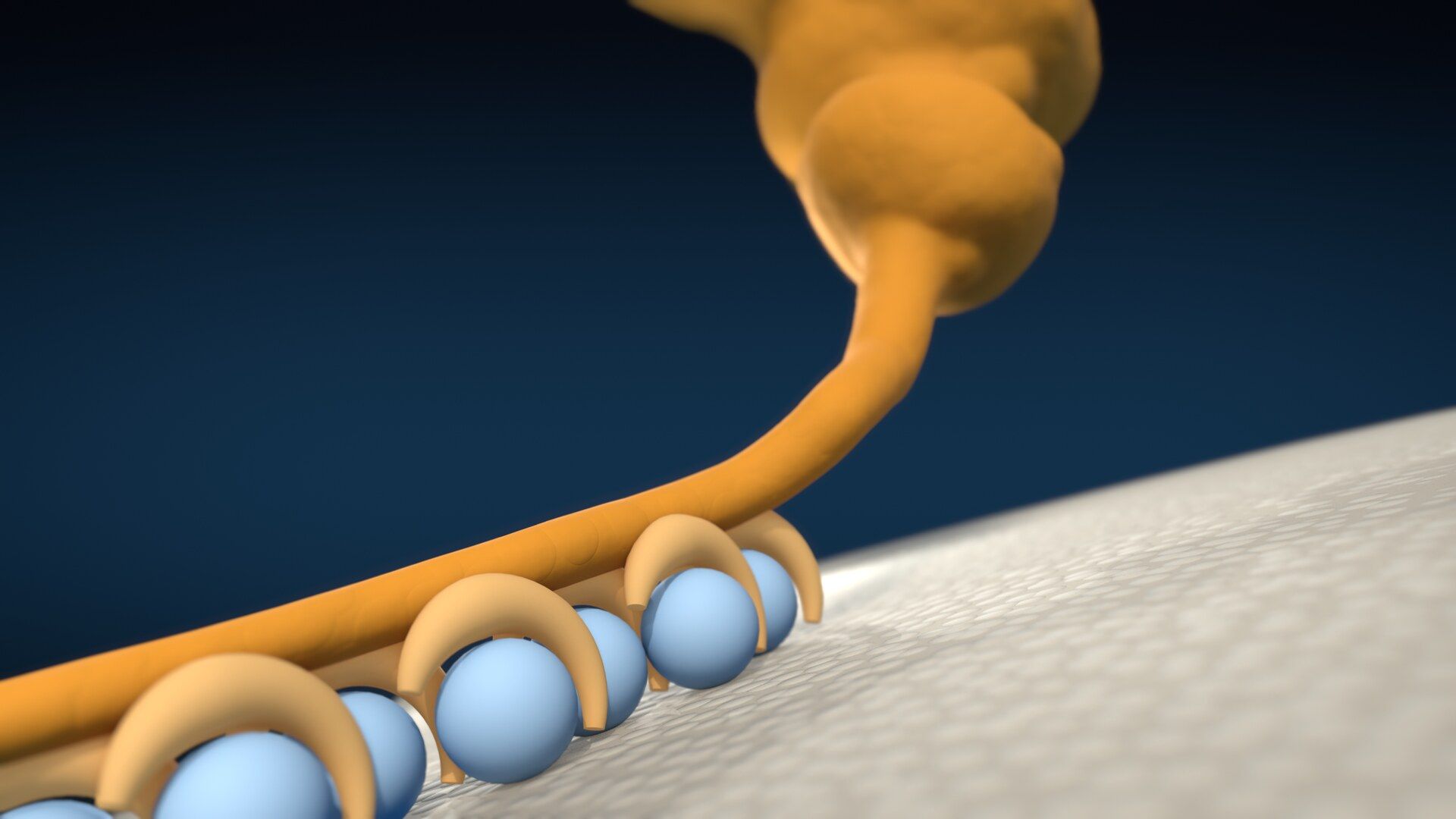
A newborn’s blood already has the proteins that make it clot. Without vitamin K, those proteins can’t do their job: the liver builds them without the hooks that let calcium settle in and the protein hold on to a platelet’s surface. Very little vitamin K crosses the placenta and breast milk carries very little, so a shortage can last for months, and the bleeding it causes can come weeks after birth.
Claims and sources
- 01
The liver builds the clotting proteins and releases them into the blood, and that part doesn’t need vitamin K. At a wound, platelets pile up to plug the gap and most of the clotting reaction then runs on their surface. To help, a clotting protein has to hold on to that surface. The unfinished ones can’t. They slide off.
“Most”, not all: the first step of clotting runs on other cells, not on platelets. The surface in the laboratory work is a phospholipid membrane in a test tube, made with a vitamin K antagonist rather than by a shortage, and tested on prothrombin. That the unfinished proteins are released is shown for the clotting factors in the picture; it is not uniform across the whole vitamin K-dependent family. “Slide off” is the picture for binding much less tightly, if at all.
PIVKAs are released from the liver into the blood but lack calcium-binding activity and are thus inactive.
Araki S, Shirahata A. Vitamin K Deficiency Bleeding in Infancy. Nutrients. 2020;12(3):780.
They provide the surface on which most of the thrombin required for clot formation is generated.
Hoffman M, Monroe DM 3rd. A cell-based model of hemostasis. Thromb Haemost. 2001;85(6):958-965.
Platelet aggregation, the process by which platelets adhere to each other at sites of vascular injury, has long been recognized as critical for hemostatic plug formation and thrombosis.
Jackson SP. The growing complexity of platelet aggregation. Blood. 2007;109(12):5087-5095.
Activated platelets expose phosphatidylserine on their outer surface, the membrane the clotting proteins bind.
Lentz BR. Exposure of platelet membrane phosphatidylserine regulates blood coagulation. Prog Lipid Res. 2003;42(5):423-438.
In comparison to normal prothrombin, the abnormal prothrombin produced in response to vitamin K antagonists has been found to bind much less tightly, if at all, to phospholipid surfaces.
Esmon CT, Suttie JW, Jackson CM. The functional significance of vitamin K action. Difference in phospholipid binding between normal and abnormal prothrombin. J Biol Chem. 1975;250(11):4095-4099.
- 02
Vitamin K drives a finishing step that adds hooks to each protein. Calcium settles into the hooks. Now the protein grips.
The hooks in the picture are a stylization: the real binding domain carries 9 to 13 of these residues, clustered at one end, and the video draws fewer. Calcium sits bound within the domain; the video’s calcium beads are a picture of that. The structural study is of a bovine protein; the surface it names is a membrane carrying phosphatidylserine, as on activated platelets. The letter K, shown on screen as a side note, is the discoverer’s own account: the next letter not yet used for a vitamin, and the first letter of “koagulation”.
Carboxylation is accomplished by the enzyme gamma glutamyl carboxylase (GGCX) which requires the propeptide-containing substrate and three co-substrates: reduced vitamin K, CO2, and O2.
Stafford DW. The vitamin K cycle. J Thromb Haemost. 2005;3(8):1873-1878.
Vitamin K plays a critical role as a cofactor of gamma-glutamyl carboxylase in the conversion of glutamic acid to gamma-carboxyglutamic acid residues in the N-terminus of vitamin K-dependent procoagulant factors and proteins C and S
Araki S, Shirahata A. Vitamin K Deficiency Bleeding in Infancy. Nutrients. 2020;12(3):780.
In a calcium-dependent interaction critical for blood coagulation, vitamin K-dependent blood coagulation proteins bind cell membranes containing phosphatidylserine via gamma-carboxyglutamic acid-rich (Gla) domains.
Huang M, Rigby AC, Morelli X, Grant MA, Huang G, Furie B, Seaton B, Furie BC. Structural basis of membrane binding by Gla domains of vitamin K-dependent proteins. Nat Struct Biol. 2003;10(9):751-756.
The letter K was the first one in the alphabet which had not, with more or less justification, been used to designate other vitamins, and it also happened to be the first letter in the word 'koagulation' according to the Scandinavian and German spelling.
Dam H. The discovery of vitamin K, its biological functions and therapeutical application. Nobel Lecture, December 12, 1946.
- 03
When vitamin K runs short, some babies start to bleed. Sometimes in the first week. Sometimes weeks later: a few in every hundred thousand, and then often inside the skull.
On screen
4 to 7 per 100,000
Late bleeding without vitamin K at birth · two European surveys
The 4.4 and 7.2 per 100,000 are the British Isles and German surveys of 1988 to 1990, taken from a review’s table (secondary) and quoted by a national health report; the denominator is infants given no vitamin K at birth. The review’s own median across surveys is far higher. Late bleeding is defined as after the first week and before six months, with a peak between 2 and 8 weeks; the share inside the skull is 30 to 60% in the review and varies by survey. Early warning bleeds are documented in some cases, so the video does not say the bleeding comes without warning.
incidence of late VKDB ranges from 4.4 to 7.2 per 100,000 infants
Warren M, Miller A, Traylor J, Sidonio R, Morad A, Goodman A, Christensen D, Grant A, Odom E, Okoroh E, Clayton J, Maenner M, Marcewicz L. Notes from the Field: Late Vitamin K Deficiency Bleeding in Infants Whose Parents Declined Vitamin K Prophylaxis — Tennessee, 2013. MMWR Morb Mortal Wkly Rep. 2013;62(45):901-902.
Without prophylaxis at birth, late bleeding was 4.4 per 100,000 in the British Isles survey and 7.2 per 100,000 in the German survey (Table 4); intracranial bleeding is the predominant manifestation of late onset bleeding.
Intracranial bleeding (30 to 60%) is the predominant manifestation of late onset VKDB,
Sankar MJ, Chandrasekaran A, Kumar P, Thukral A, Agarwal R, Paul VK. Vitamin K prophylaxis for prevention of vitamin K deficiency bleeding: a systematic review. J Perinatol. 2016;36(Suppl 1):S29-S35.
VKDB can be classified by age of onset into early (<24 h), classical (days 1-7) and late (>1 week <6 months),
Sutor AH, von Kries R, Cornelissen EA, McNinch AW, Andrew M. Vitamin K deficiency bleeding (VKDB) in infancy. ISTH Pediatric/Perinatal Subcommittee. Thromb Haemost. 1999;81(3):456-461.
In British and Irish surveillance, intracranial bleeding was among the late presentations, and late onset bleeding remained virtually confined to breast-fed infants who received either no vitamin K or one oral dose.
McNinch A, Busfield A, Tripp J. Vitamin K deficiency bleeding in Great Britain and Ireland: British Paediatric Surveillance Unit Surveys, 1993-94 and 2001-02. Arch Dis Child. 2007;92(9):759-766.
In a Japanese national survey of babies over 2 weeks old with no obvious cause, intracranial haemorrhage was seen in most of the idiopathic cases.
Hanawa Y, Maki M, Murata B, et al. The second nation-wide survey in Japan of vitamin K deficiency in infancy. Eur J Pediatr. 1988;147(5):472-477.
- 04
Very little vitamin K crosses the placenta. You’ll often hear it’s because newborns lack gut bacteria. That may play a part, but the evidence is thin. After birth the supply stays low: breast milk carries very little vitamin K, so the shortage doesn’t end at birth. It can last for months.
The placenta figure is from nine mother and baby pairs; one later study found cord levels nearer two thirds of the mother’s, so no number is spoken. The gut-bacteria review says the idea is “enticing but presently lacks hard evidence”; it says unproven, not wrong. A second review calls the bacterial supply to newborns “questionable”. A third argues the colon is not a significant source for humans generally. Not every review agrees: an older one holds that bacterial menaquinones play a significant role in humans in general, not newborns specifically. Breast milk values vary by assay; the study followed 23 infants. Formula has vitamin K added, about 50 times as much as breast milk (on screen as a side note). Formula is required by U.S. rule to carry a minimum content.
The large concentration gradient between maternal and neonatal plasma suggests that vitamin K1 does not cross the placenta readily or that the uptake by fetal plasma is low, perhaps because of low levels of a binding lipoprotein.
Shearer MJ, Rahim S, Barkhan P, Stimmler L. Plasma vitamin K1 in mothers and their newborn babies. Lancet. 1982;2(8296):460-463.
The American Academy of Pediatrics statement reports that little vitamin K crosses the placenta and that breast milk provides relatively low levels.
Hand I, Noble L, Abrams SA; AAP Committee on Fetus and Newborn, Section on Breastfeeding, Committee on Nutrition. Vitamin K and the Newborn Infant. Pediatrics. 2022;149(3):e2021056036.
The implication that low hepatic reserves of MK may be a major contributory factor to the brittleness of neonatal vitamin K status is enticing but presently lacks hard evidence.
Shearer MJ, Fu X, Booth SL. Vitamin K nutrition, metabolism, and requirements: current concepts and future research. Adv Nutr. 2012;3(2):182-195.
questionable amounts from vitamin K present in the intestinal tract synthesized from bacteria
Greer FR. Vitamin K the basics—what's new? Early Hum Dev. 2010;86 Suppl 1:43-47.
Plasma phylloquinone concentrations in the infants fed human milk remained extremely low (mean less than 0.25 ng/mL) throughout the first 6 months of life
Greer FR, Marshall S, Cherry J, Suttie JW. Vitamin K status of lactating mothers, human milk, and breast-feeding infants. Pediatrics. 1991;88(4):751-756.
phylloquinone contents of milk formulas are typically 50-fold higher than human milk
Shearer MJ, Fu X, Booth SL. Vitamin K nutrition, metabolism, and requirements: current concepts and future research. Adv Nutr. 2012;3(2):182-195.
U.S. Code of Federal Regulations, Title 21, section 107.100, Infant formula: nutrient specifications.
- 05
That’s why the bleeding can come weeks later. One dose at birth is given to cover that gap. The American Academy of Pediatrics has recommended vitamin K for all newborns since 1961.
On screen
0 to 0.25 per 100,000
Late bleeding with vitamin K injected at birth · the same two surveys
The two rates are from the same review’s table as the figures without it (secondary), with no interval shown for the German figure. The pooled comparison across surveillance studies is observational, not from trials.
With intramuscular vitamin K at birth, late bleeding was 0 per 100,000 in the British Isles survey and 0.25 per 100,000 in the German survey (Table 4).
Data from four surveillance studies indicate that the use of IM/subcutaneous vitamin K prophylaxis could significantly reduce the risk of late VKDB when compared with no prophylaxis
Sankar MJ, Chandrasekaran A, Kumar P, Thukral A, Agarwal R, Paul VK. Vitamin K prophylaxis for prevention of vitamin K deficiency bleeding: a systematic review. J Perinatol. 2016;36(Suppl 1):S29-S35.
Intramuscular administration of vitamin K for prevention of vitamin K deficiency bleeding (VKDB) has been a standard of care since the American Academy of Pediatrics recommended it in 1961.
Hand I, Noble L, Abrams SA; AAP Committee on Fetus and Newborn, Section on Breastfeeding, Committee on Nutrition. Vitamin K and the Newborn Infant. Pediatrics. 2022;149(3):e2021056036.
- 06
When babies with this type of bleeding are given vitamin K, their clotting tests normalize in a day. The proteins are there. The vitamin K helps them hold on.
On screen
normal at 24 hours
Prothrombin time after vitamin K · 120 infants
One hospital, one time point, and one test (prothrombin time). Partly true by definition: the case definition required clotting times that normalized within 12 to 24 hours of vitamin K, and the condition is itself defined as correctable by vitamin K.
PT was normalized after 24 h of administration of vitamin K in all patients, and no additional bleeding was observed.
Per H, Arslan D, Gümüş H, Çoskun A, Kumandaş S. Intracranial hemorrhages and late hemorrhagic disease associated cholestatic liver disease. Neurol Sci. 2013;34(1):51-56.
VKDB is bleeding due to inadequate activity of VK-dependent coagulation factors (II, VII, IX, X), correctable by VK replacement.
Sutor AH, von Kries R, Cornelissen EA, McNinch AW, Andrew M. Vitamin K deficiency bleeding (VKDB) in infancy. ISTH Pediatric/Perinatal Subcommittee. Thromb Haemost. 1999;81(3):456-461.
- Picture
The clotting protein is drawn as a stylization, not a solved structure; its hook end follows a structural study of the binding domain.
The protein is a bent chain of lobes with a thin tail whose end carries a cluster of hooks, each holding one calcium. The model of the hook end follows the structure from the structural study, which has 10 gamma-carboxyglutamic acid residues and 7 calcium ions; across the clotting proteins the count runs from 9 to 13, clustered in a folded domain at one end, and the picture draws 7 hooks. Calcium sits bound within the domain and its calcium-bearing face is the one that meets the membrane; there is no separate calcium layer. The structure is of a bovine prothrombin domain with a lipid, not a human clotting protein.
Huang M, Rigby AC, Morelli X, Grant MA, Huang G, Furie B, Seaton B, Furie BC. Structural basis of membrane binding by Gla domains of vitamin K-dependent proteins. Nat Struct Biol. 2003;10(9):751-756.
Left out
Warfarin, which blocks the recycling of vitamin K and so switches this same step off on purpose.
Cut for length; it arrived before the newborn question was answered.
A number for how much faster the clotting reaction runs on a platelet’s surface.
The review figures disagree (about 150,000-fold for the whole enzyme complex against 278,000 in an older study) and none isolates the membrane’s own share.
The wider range of late bleeding, 4.4 to 72 per 100,000, which includes Asian surveys.
The video uses the two European surveys that report both with and without vitamin K at birth, and says “a few in every hundred thousand”.
“81 times” more likely to bleed late without it.
It traces to a 1991 study of 27 cases, a small base, and a relative risk adds nothing the rate doesn’t already show.
Corrections
None.
Report an error: contact@worm.dev
Transcript
Here's why newborns get vitamin K. Their blood already has the proteins that make it clot. But without vitamin K, those proteins can't do their job. And when it runs short, some babies start to bleed. Sometimes in the first week. Sometimes weeks later: a few in every hundred thousand, and then often inside the skull. Why weeks later?
We need to start in the liver. It builds the clotting proteins and releases them into the blood. That part doesn't need vitamin K. At a wound, platelets pile up to plug the gap. Most of the clotting reaction then runs on their surface. To help, a clotting protein has to hold on to that surface.
The unfinished ones can't. They slide off.
Vitamin K drives a finishing step that adds hooks to each protein. Calcium settles into the hooks. Now the protein grips.
So why do newborns lack vitamin K?
Very little vitamin K crosses the placenta. You'll often hear it's because they lack gut bacteria. That may play a part, but the evidence is thin.
After birth the supply stays low. Breast milk carries very little vitamin K. So the shortage doesn't end at birth. It can last for months. That's why the bleeding can come weeks later.
One dose at birth is given to cover that gap.
The American Academy of Pediatrics has recommended vitamin K for all newborns since 1961. When babies with this type of bleeding are given vitamin K, their clotting tests normalize in a day. The proteins are there. The vitamin K helps them hold on.
Reference list
- Araki S, Shirahata A. Vitamin K Deficiency Bleeding in Infancy. Nutrients. 2020;12(3):780. doi.org/10.3390/nu12030780
- Hoffman M, Monroe DM 3rd. A cell-based model of hemostasis. Thromb Haemost. 2001;85(6):958-965. pubmed.ncbi.nlm.nih.gov/11434702
- Jackson SP. The growing complexity of platelet aggregation. Blood. 2007;109(12):5087-5095. doi.org/10.1182/blood-2006-12-027698
- Lentz BR. Exposure of platelet membrane phosphatidylserine regulates blood coagulation. Prog Lipid Res. 2003;42(5):423-438. doi.org/10.1016/s0163-7827(03)00025-0
- Esmon CT, Suttie JW, Jackson CM. The functional significance of vitamin K action. Difference in phospholipid binding between normal and abnormal prothrombin. J Biol Chem. 1975;250(11):4095-4099. pubmed.ncbi.nlm.nih.gov/1126944
- Stafford DW. The vitamin K cycle. J Thromb Haemost. 2005;3(8):1873-1878. doi.org/10.1111/j.1538-7836.2005.01419.x
- Huang M, Rigby AC, Morelli X, Grant MA, Huang G, Furie B, Seaton B, Furie BC. Structural basis of membrane binding by Gla domains of vitamin K-dependent proteins. Nat Struct Biol. 2003;10(9):751-756. doi.org/10.1038/nsb971
- Dam H. The discovery of vitamin K, its biological functions and therapeutical application. Nobel Lecture, December 12, 1946. nobelprize.org/uploads/2018/06/dam-lecture.pdf
- Warren M, Miller A, Traylor J, Sidonio R, Morad A, Goodman A, Christensen D, Grant A, Odom E, Okoroh E, Clayton J, Maenner M, Marcewicz L. Notes from the Field: Late Vitamin K Deficiency Bleeding in Infants Whose Parents Declined Vitamin K Prophylaxis — Tennessee, 2013. MMWR Morb Mortal Wkly Rep. 2013;62(45):901-902. cdc.gov/mmwr/preview/mmwrhtml/mm6245a4.htm
- Sankar MJ, Chandrasekaran A, Kumar P, Thukral A, Agarwal R, Paul VK. Vitamin K prophylaxis for prevention of vitamin K deficiency bleeding: a systematic review. J Perinatol. 2016;36(Suppl 1):S29-S35. doi.org/10.1038/jp.2016.30
- Sutor AH, von Kries R, Cornelissen EA, McNinch AW, Andrew M. Vitamin K deficiency bleeding (VKDB) in infancy. ISTH Pediatric/Perinatal Subcommittee. Thromb Haemost. 1999;81(3):456-461. pubmed.ncbi.nlm.nih.gov/10102477
- McNinch A, Busfield A, Tripp J. Vitamin K deficiency bleeding in Great Britain and Ireland: British Paediatric Surveillance Unit Surveys, 1993-94 and 2001-02. Arch Dis Child. 2007;92(9):759-766. doi.org/10.1136/adc.2006.104752
- Hanawa Y, Maki M, Murata B, et al. The second nation-wide survey in Japan of vitamin K deficiency in infancy. Eur J Pediatr. 1988;147(5):472-477. doi.org/10.1007/BF00441969
- Shearer MJ, Rahim S, Barkhan P, Stimmler L. Plasma vitamin K1 in mothers and their newborn babies. Lancet. 1982;2(8296):460-463. doi.org/10.1016/s0140-6736(82)90493-7
- Hand I, Noble L, Abrams SA; AAP Committee on Fetus and Newborn, Section on Breastfeeding, Committee on Nutrition. Vitamin K and the Newborn Infant. Pediatrics. 2022;149(3):e2021056036. doi.org/10.1542/peds.2021-056036
- Shearer MJ, Fu X, Booth SL. Vitamin K nutrition, metabolism, and requirements: current concepts and future research. Adv Nutr. 2012;3(2):182-195. doi.org/10.3945/an.111.001800
- Greer FR. Vitamin K the basics—what's new? Early Hum Dev. 2010;86 Suppl 1:43-47. doi.org/10.1016/j.earlhumdev.2010.01.015
- Greer FR, Marshall S, Cherry J, Suttie JW. Vitamin K status of lactating mothers, human milk, and breast-feeding infants. Pediatrics. 1991;88(4):751-756. pubmed.ncbi.nlm.nih.gov/1896278
- U.S. Code of Federal Regulations, Title 21, section 107.100, Infant formula: nutrient specifications. ecfr.gov/current/title-21/section-107.100
- Per H, Arslan D, Gümüş H, Çoskun A, Kumandaş S. Intracranial hemorrhages and late hemorrhagic disease associated cholestatic liver disease. Neurol Sci. 2013;34(1):51-56. doi.org/10.1007/s10072-012-0965-5